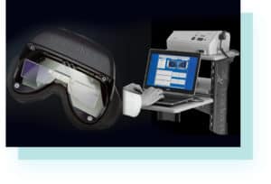
As audiology continues to evolve, new technologies are being developed to better diagnose and treat various ear and balance disorders. One such technology is videonystagmography, a non-invasive test that evaluates the functionality of the inner ear and its connection to the brain. This diagnostic tool has become an essential component of audiology assessments, allowing healthcare professionals to accurately diagnose and treat a range of conditions.
What is a Videonystagmography Test
Videonystagmography (VNG) is a diagnostic test that evaluates the vestibular system, which is responsible for maintaining balance and spatial orientation. The test utilizes infrared cameras to record eye movements and assess how the eyes respond to various stimuli, such as changes in head position or temperature.
During a VNG test, the patient wears special goggles that are equipped with infrared cameras. The cameras track eye movements and record them as the patient performs a series of movements, such as looking left and right, up and down, and tilting the head in different directions. The test also includes caloric stimulation, which involves irrigating the ear canal with warm and cold water to evaluate how the vestibular system responds to temperature changes.
One of the key benefits of VNG is that it provides a comprehensive assessment of the vestibular system, allowing audiologists to identify the root cause of balance disorders and related symptoms such as dizziness, vertigo, and nausea. The VNG testing can also help diagnose other conditions, such as acoustic neuroma, which is a benign tumor that affects the vestibular nerve.
In addition to diagnosis, VNG can also be used to monitor the progress of treatment for balance disorders. By comparing the results of multiple VNG tests over time, audiologists can determine whether a patient’s condition is improving or worsening, and adjust treatment plans accordingly.
What to Expect During a Videonystagmography Test
Before the test begins, the patient will be asked to remove any contact lenses or glasses and to avoid caffeine, alcohol, and certain medications that can affect the vestibular system. They will also undergo a physical examination and provide a detailed medical history. This information can help the audiologist determine the underlying cause of the patient’s symptoms and customize the VNG test accordingly. The test typically takes about 90 minutes to complete and is performed in a quiet, dimly lit room.
During the VNG test, the audiologist will use specialized equipment to monitor the patient’s eye movements. This equipment typically includes infrared video cameras and sensors that are attached to the patient’s forehead and around the eyes. The video cameras capture images of the patient’s eyes as they perform various tasks and movements, while the sensors measure the electrical activity in the muscles surrounding the eyes.
The VNG test typically consists of four main parts: gaze testing, tracking testing, positional testing, and caloric stimulation. During gaze testing, the patient will be asked to look at a fixed point while the audiologist moves a light or other visual target in different directions. Tracking testing involves following a moving visual target with the eyes, while positional testing evaluates how the patient’s symptoms change when they move their head or body into different positions.
The caloric stimulation portion of the VNG test involves irrigating the ear canal with warm and cold water. This can help to evaluate the function of the vestibular system on each side of the body. The patient may experience a brief sensation of vertigo or dizziness during this part of the test, but it typically subsides quickly.
After the VNG test is complete, the audiologist will review the results and provide a diagnosis. Depending on the underlying cause of the patient’s symptoms, treatment options may include medications, vestibular rehabilitation therapy, or surgery.
Overall, videonystagmography test is generally considered safe and noninvasive, with minimal side effects. However, some patients may experience mild discomfort or temporary symptoms during or after the test. Here are some potential side effects of videonystagmography test:
- Dizziness or vertigo. During the caloric stimulation portion of the VNG test, the patient’s inner ear is irrigated with warm and cold water. This can cause a brief sensation of vertigo or dizziness, but it typically subsides quickly.
- Eye irritation. The electrodes and sensors used during the VNG test may cause mild irritation or discomfort around the eyes, but this is usually temporary.
- Nausea. Some patients may experience nausea or vomiting during the test, especially if they have a history of motion sickness or inner ear problems.
- Fatigue. The VNG test can be tiring for some patients, as it requires them to remain still and focused for an extended period of time.
- Headache. Rarely, patients may experience a headache or migraine after the VNG test. This may be related to the bright lights or other sensory stimuli used during the test.
If you experience any persistent or severe symptoms after the VNG test, such as severe dizziness, headache, or vision changes, be sure to contact your doctor right away. However, most patients can resume their normal activities immediately after the test and do not experience any lasting side effects.
Interpreting VNG Results
After the VNG test is complete, the audiologist will review the results to determine if there are any abnormalities in the patient’s vestibular system. These results are presented in a series of graphs and charts that show the patient’s eye movements and responses to various stimuli.
One of the key indicators that audiologists look for in VNG results is the presence of nystagmus, which is a rapid, involuntary movement of the eyes. Depending on the direction and intensity of the nystagmus, audiologists can determine which part of the vestibular system is affected and the severity of the patient’s condition.
Other indicators that may be present in VNG results include a reduced response to caloric stimulation, which can indicate damage to the vestibular nerve, or abnormal eye movements during positional changes, which can suggest a problem with the inner ear.
Interpreting VNG results requires specialized training and expertise, as the graphs and charts can be complex and difficult to interpret without proper training. It’s important for patients to work closely with their audiologist to fully understand their VNG results and what they mean for their diagnosis and treatment plan.


